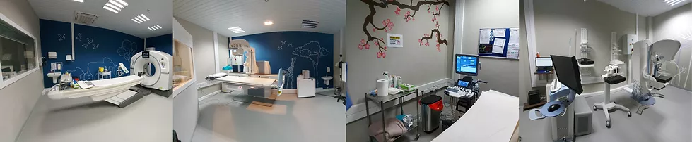
Mammography
Quest Medical Imaging in Accra, 3D Mammograms with Tomosynthesis .
2D mammograms are the regular standard procedure. Simply put it means that the breast image is obtained from top to bottom or from side to side. Visualisation of small cancers can be missed and often result in the patient having to experience further imaging and biopsies.
However, with advances in breast imaging, the early detection of breast cancer has improved by 40%. Tomosynthesis or 3D Digital Breast Imaging (DGB) is fast becoming the gold standard for imaging breast tissue, especially for women with dense breast tissue. 3D imaging has undoubtedly improved the peace of mind of both radiologists and patients.
Tomosynthesis (3D) technology is performed with the tomosynthesis mammogram machine. The software enables the x-ray tube to ‘sweep’ over the breast in a short arc movement from different angles, allowing for a layered or sliced breast tissue acquisition of images.
The Radiologist is then able to view these layers one by one. This allows for overlying structures to be visualised individually and provides clarity in those difficult, dense areas within the breast.
The advantages of Tomosynthesis are:
- Improves visualisation and detection of breast cancer.
- Fewer recalls for patients, especially regarding further biopsies. The reason is that tissue is easier to see with overlying structures. As a result, more certainty in the evaluation of tissue.
- Potential less compression of the breast due to increased visibility.

With access to