Ultrasound
An ultrasound examination uses high-frequency sound waves to create an image. The sound waves reflect off body structures such as the liver, muscles or the unborn foetus.
An image is then created by using software with a special computer. No radiation (such as is used with x-ray images) is used during an ultrasound examination.
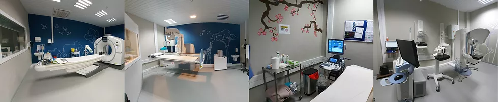
The test is a painless procedure and involves the patient lying down or sitting next to the ultrasound machine. A water-based gel is applied to the skin over the area of concern.
The gel helps to transmit the sound waves. A transducer or probe is then moved over the examination area. The ultrasound department at QMI covers general abdominal, vascular ultrasound, neonatal and paediatrics, muscular skeletal ultrasound and obstetrics and Musculo-skeletal.