CT – Computed Tomograpy
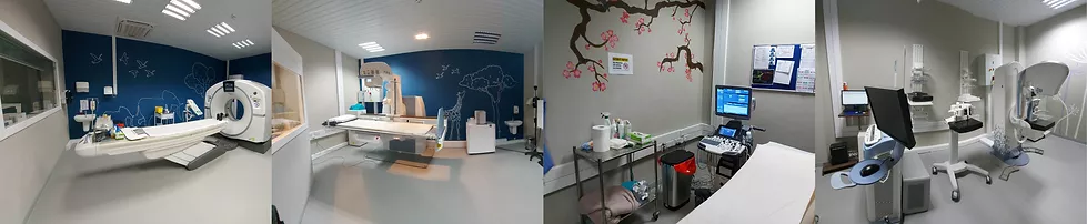
A CT scanner creates its image by moving the human body through a donut shaped machine, which emits a series of narrow x-ray beams. The data from these beams is converted by a computer into diagnostic cross- sectional images. Special software is used to reconstruct images in different planes and to create 3-dimensional views.
A CT scan provides fast and detailed imaging of the head and the body. CT is particularly useful in imaging the abdomen and pelvis, the lungs, soft tissues, bones, brain, heart and blood vessels.
Because of the speed of the examination, it is possible to image the blood vessels in the brain, neck, lungs, Abdomen, upper and lower extremities. Most CT scans are painless and non-invasive.
Some examinations require special preparations and an intravenously administered contrast (dye) media to optimize the visualization of organs, structures and vessels in the body. Patients are screened for known allergies and existing conditions before any procedure is started.